buy online

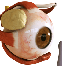
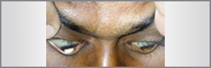 Often appearing in the teens or early twenties, keratoconus is a progressive disease in which the normally round cornea thins and begins to bulge into a cone-like shape. This cone shape deflects light as it enters the eye on its way to the light-sensitive retina, causing distorted vision. Keratoconus can occur in one or both eyes.
Signs and Symptoms of Keratoconus
- Astigmatism
- Eye rubbing
- Nearsightedness
- Ghost images
- Glare at night
- Light sensitivity
- Blurred vision even when wearing glasses and contact lenses
- Frequent prescription changes in glasses and contact lenses
Causes for Keratoconus
 New research has demonstrated a relationship between malfunction of beneficial enzymes found within the eye’s surface and an accompanying chemical imbalance that leads to toxic damage and thinning of eye tissue. Because keratoconus can be found in extended families, this improper functioning of beneficial enzymes appears to have genetic causes about 5% of the time. Eye damage from keratoconus also can be linked to factors such as overexposure to sunlight, improper fittings of contact lenses, excessive eye rubbing, and continual (chronic) eye irritation.
How Keratoconus Is Detected and Diagnosed?
Keratoconus is usually diagnosed when patients reach their 20’s. For some, it may advance over several decades, for others, the progression may reach a certain point and stop. Keratoconus is not usually visible to the naked eye until the later stages of the disease. In severe cases, the cone shape is visible to an observer when the patient looks down while the upper lid is lifted. When looking down, the lower lid is no longer shaped like an arc, but bows outward around the pointed cornea. This is called Munson’s sign.
Special corneal testing called topography provides the doctor with detail about the cornea’s shape and is used to detect and monitor the progression of the disease. A pachymeter may also be used to measure the thickness of the cornea.
Treatment for Keratoconus
 The first line of treatment for patients with keratoconus is to fit rigid gas permeable (RGP) contact lenses. Because this type of contact is not flexible, it creates a smooth, evenly shaped surface to see through. However, because of the cornea’s irregular shape, these lenses can be very challenging to fit. This process often requires a great deal of time and patience. When vision deteriorates to the point that contact lenses no longer provide satisfactory vision, corneal transplant may be necessary to replace the diseased cornea with a healthy one.
Exclusive Treatment Options at Shekhar Eye Research Centre
| Chandra Scleral Lenses | Specialty RGP Lenses |
| Rose K Contact Lenses | Riboflavin |
| Intacts | Deep Lammelar Keratoplasty |
| Penetrating Keratoplasty |
 OUR SPECIALIST

Mr. Chandrashekhar Chawan , is an Optometrist, Ocularist and Ocular scientist. He was the first in the world to develop Soft Hydrogel Ocular Prosthesis in 1999.
He has many first to his credits, recently he has ..
.jpg)
Mr. Chandrashekhar Chawan , is an Optometrist, Ocularist and Ocular scientist. He was the first in the world to develop Soft Hydrogel Ocular Prosthesis in 1999.
He has many first to his credits, recently he has ..
.jpg)
Mr. Chandrashekhar Chawan , is an Optometrist, Ocularist and Ocular scientist. He was the first in the world to develop Soft Hydrogel Ocular Prosthesis in 1999.
He has many first to his credits, recently he has ..
 our products
- 3D Custom Made Artificial Eyes
- Keratoconus
- Oculoplasty
- Orthokeratology
- Speciality Contact Lens
- Chandra Scleral Lenses
- Myopia Control
- Gold Contact Lenses
 phone
phone
 email
email

Copyright ©
Shekhar eye. All rights reserved108 Falcon Court,
Hari Om Nagar, Mulund (E),
Mumbai 400081 ( India)